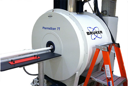

Bruker Pharmascan 70/16 7T MRI scanning system
The Bruker Pharmascan MRI system is a state of the art small animal MRI unit that consists of a 7 Tesla magnet, the Bruker Avance III spectrometer that provides direct digitalization of radio frequency signals, imaging gradients that provide up to 370 mTesla/meter gradient strength, and a collection of imaging coils. The largest coil has a 7.2 cm inner diameter into which the subject/specimen must fit. The system is equipped with full physiologic support for small animals during scans with thermal support and real time display of rectal temperature, respiration and ECG waveforms.
The Visualsonics VEVO 2100 ultrasound system is equipped with the MS250 and MS550S probes. The system has capabilities to perform high resolution cardiac and abdominal scans in small animals, as well as to guide deep needle injections. Installed modules include B mode, M mode, color doppler mode, pulse wave (PW) doppler mode, tissue doppler mode, and contrast mode. More details can be found here.
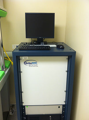
EchoMRI-100V whole body magnetic resonance analyzer
Echo MRI is a versatile tool for preclinical studies associated with nutrition and growth. Echo MRI provides measurement of whole body fat, free water, lean tissue water and total water masses in mice based upon NMR relation rates. Measurements are obtained from mice without need for anesthesia and are typically completed within approximately 5-7 minutes per animal.
A computer system comprised of a Mac Pro computer, a Mac mini computer, a Macbook Air computer, a Synology DS1813+ expandable network attached storage (NAS) system with 6-4Terabyte disk cartridges, and network router and switches for offline data archival and analysis. Two Harvard apparatus forced air ventilators (mouse and rat), two isoflurane mixers to provide anesthesia during imaging, several recirculated heated water pumps with pads to provide thermal support of animals during imaging. A network analyzer for testing/analyzing radiofrequency MRI coils.
GE eXplore Locus CT scanner system
The Trifoil eXplore RS9 microCT system is a cone-beam system that operates at up to 80 kVp and 450 uAmp. This system provides CT imaging of specimens and small animals within a field of view of 6.0 cm radial and 4.0 cm axial at resolutions of approximately 0.27, 0.48, or 0.97 mm in 3 dimensions. MicroCT is useful for measuring general anatomy, bone structure, and bone density.
Visualsonics VEVO 2100 Ultrasound
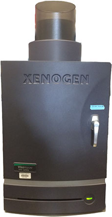
IVIS Lumina
The fluorescent and bioluminescent imaging system, provides innovative ways for imaging biochemical process in small animal models. It is useful for studying therapeutic responses of tumor to various therapies, tracer distribution analysis. IVIS lumina has extended capability to function in the infra-red region of the visible electromagnetic spectrum.
Work Station and Data Storage