
Bruker Pharmascan 7T MRI system - capabilities
The Bruker MRI system capabilities include a full suite of MRI pulse sequences and numerous applications such as contrast enhanced imaging, respiration and/or cardiac gated imaging, diffusion weighted imaging, diffusion tensor mapping, NMR relaxation rate mapping, fMRI, angiography, flow velocity mapping in arteries/veins, magnetization transfer imaging, and T1rho mapping, among other methods.
Application Examples
Cardiac MRI
Cardiac MRI is typically performed with scan acquisition triggered by ECG and respiratory waveforms.


Cine
X axis tags
Y axis tags
combined
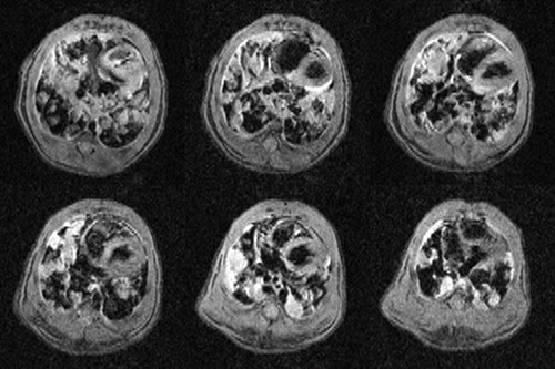
CINE MRI is a gradient echo method that provides images of multiple phases of the cardiac cycle at each slice location from apex to base of the heart in short axis view. Typically 12-18 frames during the cardiac cycle is possible, depending on heart rate. By morphometry of end diastolic and an end systolic frames of all slices of the heart one may obtain a number of metrics of cardiac perfomance, including end systolic and end diastolic volumes, ejection fraction, cardiac output, and circumferential wall thickening.
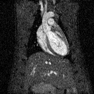
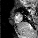
Cardiac tagging. Radiofrequency excitation applied simultaneously with gradient pulses prior to MRI readout can be used to lay out an array of parallel null lines across the image of stationary regions. In regions that move during the time between RF excitation and image readout the tag lines bend according to the displacement.
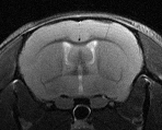
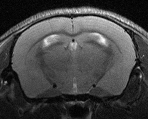
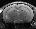
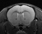
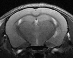
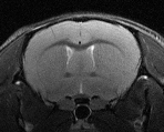
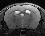
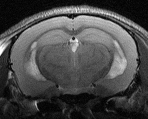
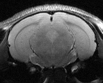
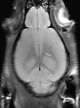
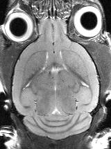
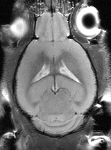
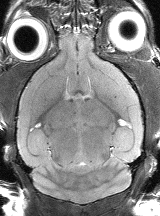
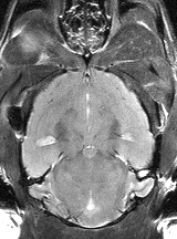
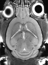
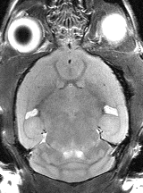
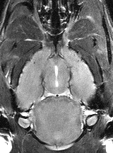
Mouse Brain MRI
Coronal view
Axial view
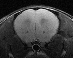


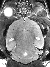
Mouse lung tumor model